Case 13 Part 10
Cirrhosis.
What’s inside?
1. Coronary ligament
2. Liver, bare area
3. Falciform ligament
4. Liver, right lobe
5. Coronary ligament
6. Liver, bare area
7. Falciform ligament
8. Liver, right lobe
9. Gallbladder
10. Liver, caudate lobe
11. Portion of diaphragm overlying liver (right lobe)
12. Gallbladder
13. Liver, quadrate lobe
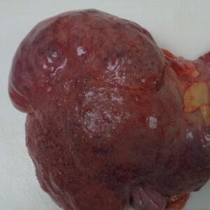
14. Liver, cirrhotic nodule
15. Liver, capsule
16. Liver, cirrhotic scarring
17. Hepatic vein
18. Spleen
19. Splenic parenchyma (white pulp is difficult to identify due to decomposition).
20. Splenic capsule
21. Panreas, head
22. Pancreas, tail
23. Pancreas, body
24. Pancreas, saponification (postmortem change)
25. Kidney
26. Kidney, deep cortical scar
27. Kidney, pelvis
28. Kidney, cortex
29. Kidney, medulla (medullary pyramid)
30. Renal pelvis (mucosa)
31. Renal pelvic fat
32. Testis, head of epididymis
33. Testis, tunica albuginea, external
34. Testis, seminiferous tubules
35. Testis, tunic albuginea
36. Bladder wall
37. Bladder mucosa
38. Bladder mucosa overlying prostate
39. Prostate, left lobe, anterior view
40. Urethra
41. Prostate, right lobe, anterior view
42. Esophagus, muscosa
43. Gastroesophageal junction
44. Stomach, body
45. Stomach, pylorus
46. Esophagus, mucosa
47. Gastroesophageal junction
48. Gastric mucosa
49. Small intestine, mucosa
50 Small intestine
51. Cecum, mucosa
52. Ascending colon, mucosa
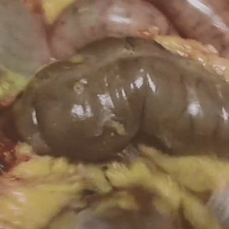
53. Two (2) polyps in mucosa of transverse colon
54. Diverticula in rectosigmoid colon
55. Two (2) polyps in mucosa of transverse colon
56. Diverticula
57. Adrenal (thinned, cortext and medulla are difficult to discern; bright yellow patches are cortical tissue)
1. Coronary ligament
2. Liver, bare area
3. Falciform ligament
4. Liver, right lobe
5. Coronary ligament
6. Liver, bare area
7. Falciform ligament
8. Liver, right lobe
9. Gallbladder
10. Liver, caudate lobe
11. Portion of diaphragm overlying liver (right lobe)
12. Gallbladder
13. Liver, quadrate lobe
14. Liver, cirrhotic nodule
15. Liver, capsule
16. Liver, cirrhotic scarring
17. Hepatic vein
18. Spleen
19. Splenic parenchyma (white pulp is difficult to identify due to decomposition).
20. Splenic capsule
21. Panreas, head
22. Pancreas, tail
23. Pancreas, body
24. Pancreas, saponification (postmortem change)
25. Kidney
26. Kidney, deep cortical scar
27. Kidney, pelvis
28. Kidney, cortex
29. Kidney, medulla (medullary pyramid)
30. Renal pelvis (mucosa)
31. Renal pelvic fat
32. Testis, head of epididymis
33. Testis, tunica albuginea, external
34. Testis, seminiferous tubules
35. Testis, tunic albuginea
36. Bladder wall
37. Bladder mucosa
38. Bladder mucosa overlying prostate
39. Prostate, left lobe, anterior view
40. Urethra
41. Prostate, right lobe, anterior view
42. Esophagus, muscosa
43. Gastroesophageal junction
44. Stomach, body
45. Stomach, pylorus
46. Esophagus, mucosa
47. Gastroesophageal junction
48. Gastric mucosa
49. Small intestine, mucosa
50 Small intestine
51. Cecum, mucosa
52. Ascending colon, mucosa
53. Two (2) polyps in mucosa of transverse colon
54. Diverticula in rectosigmoid colon
55. Two (2) polyps in mucosa of transverse colon
56. Diverticula
57. Adrenal (thinned, cortext and medulla are difficult to discern; bright yellow patches are cortical tissue)
Liver
cirrhosis
cirrhotic
nodule
gallbladder
spleen
red pulp
capsule
pancreas
saponification
adrenal
kidney
pelvis
renal pelvis
pelvic fat
urothelium
mucosa
cortex
medulla
pyramid
prostate
testes
tunica albuginea
seminiferous tubules
intestine
colon
small intestine
large intestine
polyp
diverticulum
diverticula
stomach
stomach contents
undigested digested gastric contents
esophagus
esophageal mucosa
gastric mucosa
gastroesophageal junction
cirrhosis
cirrhotic nodule
bladder
bladder mucosa